Isotropic Generalized Diffusion Tensor MRI
Scientists at the Eunice Kennedy Shriver National Institute for Child Health and Human Development (NICHD) have developed a method implemented as pulse sequences and software to be used with magnetic resonance imaging (MRI) scanners and systems. This technology is available for licensing and commercial development. The method allows for measuring and mapping features of the bulk or average apparent diffusion coefficient (ADC) of water in tissue – aiding in stroke diagnosis and cancer therapy assessment. The pulse sequences and software enable MRI scanners to yield diffusion weighted images (DWI) that are orientationally averaged. The resulting bulk diffusion signals are describable by second and higher-order tensors (HOT). This is most notably beneficial for resolving intrinsic differences in tissue water diffusivities between white and gray matter.
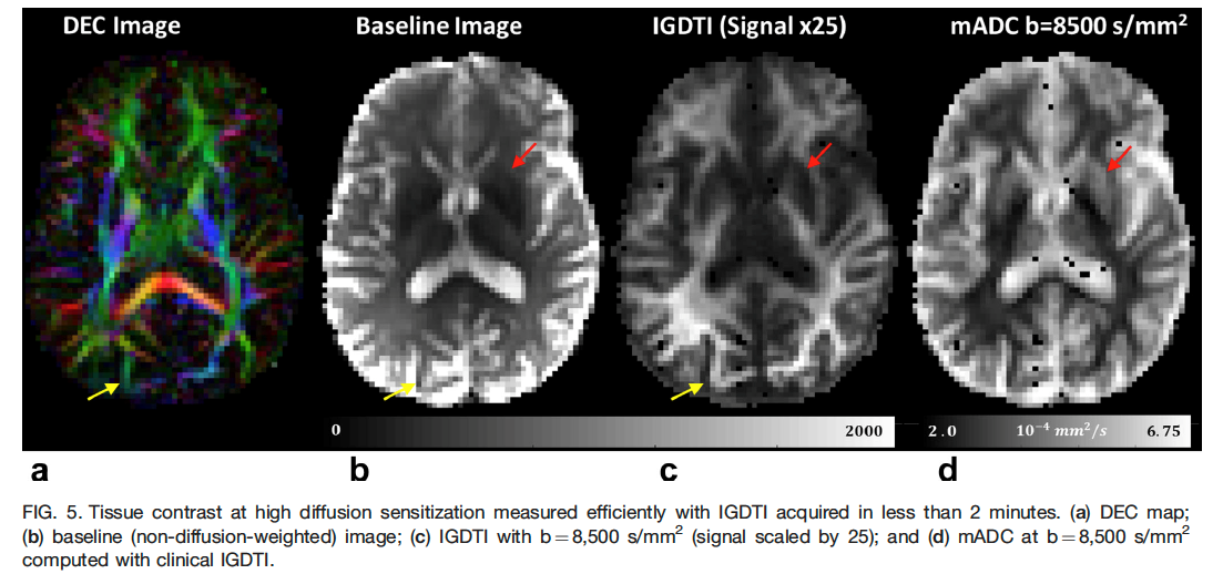
The mean apparent diffusion coefficient (mADC), or the Trace of the 2nd-order diffusion tensor, has revolutionized the diagnosis of stroke and cancer. It quantifies orientationally-averaged bulk properties of tissue water in the central nervous system while removing or averaging over the orientational bias in diffusion within white matter. However, mADC use has been limited to low diffusion sensitization. At higher diffusion sensitization (as measured by b-values substantially greater than 1000 s/mm2), DWI signal modulations due to diffusion anisotropy becomes more pronounced, especially in regions with complex microstructure, confounding the clinical evaluation. This invention overcomes this limitation by extending the range of diffusion sensitization or b-values producing DWIs that are isotropically weighted.
The technology, called isotropic-diffusometry imaging or isotropic generalized diffusion tensor imaging (IGDTI), permits measurements within clinically feasible scan times of orientationally averaged DWIs and mADCs over a wide range of b-values, and estimation of various rotation-invariant microstructural parameters, such as HOT-Traces or mean t-kurtosis, as well as estimation of the probability density function or spectrum of orientationally averaged mADCs, p(mADC), as a function of the isotropic mADC, within each imaging voxel. It also furnishes estimates of the signal fractions of various tissue types based on tissue-specific isotropic diffusivity spectra. The method selects sets of applied magnetic field gradient magnitudes and directions whose averages produce mADCs over a wide range of b-values especially those required to measure HOTs. These experimental designs are based on analytical descriptions of the tensors and can be combined to produce mADCs or probability density functions of intravoxel mADC distributions (figure below).

Competitive Advantages:
- Enables diffusion MRI methods that estimate biologically-specific rotation-invariant parameters for quantifying tissue water motilities or other intrinsic tissue characteristics within clinically feasible scan durations
- Suitable for imaging moving tissue, such as whole-body imaging, including fetal and placental MRI application
- Offers greater dynamic b-value range than other DWI based methods
- Can provide sub-voxel resolution, revealing distinct water compartments
- Enhances tissue segmentation, clustering and classification applications
- Expected to be useful in assessing a variety biological processes, pathologies, disorders and diseases
Commercial Applications:
- Potential diagnosis of disorders in the CNS and PNS, e.g., stroke, Amyotrophic lateral sclerosis (ALS), Alzheimer’s disease, Traumatic brain injury (TBI)/concussion, Cancer, Multiple sclerosis (MS)
- Incorporation within a “suite” of neuro and whole-body radiology packages
- Measuring and mapping new quantitative imaging biomarkers
- Potentially repurposing MR data already used to produce and implement other advanced diffusion MRI applications, such as Mean Apparent Propagator (MAP) MRI, Diffusion Kurtosis Imaging (DKI), or diffusion spectrum imaging (DSI)
